i foraminiferi

martedì 16 novembre 2021

A very special bacterium

 A very special bacterium

 Bacteria are unicellular procaryotic organisms. They are very small in size, too small to be seen with the naked eye, and have a very simple structure. In general the word “bacteria” is evocative of diseases and dangers. The fact that not all bacteria are dangerous and that, on the contrary, we could not leave without bacteria will be the subject of  an additional post. Here I want only to introduce a very special bacterium we discovered in our laboratory at Pisa University.
Like the majority of bacteria it is small (oval in shape 2 µm, that is two thousandths of millimiter in length and 1 µm in width) but possesses a complex structure and is far for being dangerous! We called this bacterium “Epixenosome”  (from the ancient Greek external alien body) because it lives on the dorsal surface of ciliated protozoa of Euplotidium genus.
Why epixenosomes are so special?
First of all because, as you can see in the picture, their structure is more complicated than that of the majority of known bacteria. Some characteristcs are more eukaryotic than prokaryotic and a  functional compartmentalization corresponds to the structural complexity,

Longitudinal section of epixenosomes (transmission electron microscope)

  Their DNA is localized in the upper region, is bound to basic proteins and,
at the electron microscope, it assumes a chromatin-like appearance. Moreover in the cytoplasm there are bundles of tubules in all  likelihood consisting of tubulin (a protein that up to now was considered only eukaryotic).  But the more astonishing structure is, in my opinion, the extrusive apparatus.
 The extrusive apparatus is a very well engineered structure.  It consists of a ribbon rolled up around a central core. In resting position it is compact, with shape and size well adapted to that of the intact epixenosome. The detection of external signals through membrane receptors starts up the extrusive process.
During the ejection  the ribbon unrolls from the inside by the slipping of the layers one into the other (like when we make a nose with streamers). Thus a tube forms which passes through the opening of the cell membrane (the first step of the ejecting process), and takes away the apical portion of the epixenosome containing  the genetic material.  At the end of the process the tube is 40 mm long,  that is 20 times the length of the organism, with a head 2mm long. The wall of the tube consists of the layers that are rolled up in the resting state but which are now extended, one after the other, with oblique overlapping thus ensuring the continuity of the tube.  At present we do not know the meaning of the ejection, which causes the dispersion of the epixenosomal DNA, for the bacterium itself.


 The tube at the end of the ejection

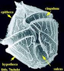
Euplotidium with some ejected epixenosomes
We know however that the ejection of epixenosomes is useful for the ciliate host. Indeed we experimentally demonstrated that predator ciliates do not eat Euplotidium with epixenosomes while easily eat those without. Note that Euplodidium without epixenosomes have never been found in nature: they can only been obtained in the laboratory. This means that  the presence of epixenosomes and their ejection is not vital for the host but represents an efficient defensive tool in nature. 
Other ciliates are able to escape predators by means of their own extrusomes:  ciliates of Euplotidium genus grow epixenosomes on their surface for their defence. For this reason in an american blog they have been compared to James Bond, the 007 agent able to invent different special weapons!


venerdì 7 maggio 2021

DINOFLAGELLATE PECULIARITIES


My posts on eukaryotic microorganisms  (protists) mostly concern with Ciliates the group I know better. I studied them for more than 40 years! However there are other groups worthy of mention for their notable peculiarities.

I already mentioned Flagellates  characterized by the possession of one or more flagella, which are long, tapering, hair-like appendages that act as organelles of locomotion. Among Flagellates a particularly interesting group are certainly Dinoflagellates  that are mostly part of the marine plankton, but also can be found in freshwater habitats.  They typically posses two flagella: a transverse flagellum running along an equatorial sulcus that beats to the cell's left, and a longitudinal flagellum, that beats posteriorly. Most are covered by a shell called lorica  often consisting of cellulose plates. They greatly differ from each other in shape and their size varies from 8 to 500 micrometers.


Lorica of a Dinoflagellate 


Ceratium

Many are autotrophs, i.e. they can produce its own food using light, water, carbon dioxide, or other chemicals thus, like plants, they are primary producer in the food chain.

Others are instead hetrotrophs so they need to eat.

Many of them are extremely voracious predators able to ingest very large preys. 

The species covered by a rigid lorica possess specific devices to capture and gulp down food organisms. Some  draw prey to the sulcal region of the cell (either via water currents set up by the flagella or via pseudopodial extensions) and ingest the prey through the sulcus.  Other species extrude a large feeding veil — a pseudopod called the pallium - to capture prey which is subsequently digested extracellularly  or  feed by attaching to its prey and ingesting prey cytoplasm through an extensible peduncle.


Dinophysis acuminata. Light micrographs of live cells feeding on a ciliate 

 Preys, attracted through chemical stimuli, are protists or even small  hurt metazoa. The latter can be co-digested by  a number of  predators.

Most planktonic dinoflagellates perform diurnal vertical migrations. In some cases the behaviour pattern is for the cells to migrate downwards away from the surface as the light becomes less intense and then to ascend again at the sunrise. In these migrations they  cover up to 50 meters.

Considering their size a distance corresponding to 4000 km for a human being!!!!

      The significance of this migration is understandable in autotrophic species needing light for their metabolism.

         But also a number of heterotophic species behave in the same way. Why?

The term plankton is a collective name for all marine and freshwater organisms nonmotile or too small or weak to swim against the current, existing in a drifting state. The term includes certain protists among which Dinoflagellates, bacteria, crustaceans, molluscs and coelenterates interacting in different ways; in particular prey-predators interactions influence reciprocal behaviours. Thus while small crustaceans, which are the main predators  of autotrophic or heterotrophic Dinoflagellates, migrate in the deepwaters during the day to prevent their own ingestion  from fishes and return to the surface in the night, it is possible that 
 Dinoflagellates migrate in the opposite sense to escape their engulfement.

 Anyway Dinoflagellate migration is a very important phenomenon in the planktonic ecosystem because it creates a massive biomass and nutrients shifting in the water column.

Dinoflagellates are the main eukaryotic protists that are capable of producing light. Within this group, bioluminescence is present in a number of ecologically important species, many of which like Noctiluca form blooms. When the organisms perceive the presence of a predator like for example a little crustacean, emit a flash of cold light lasting 0.1-0.5 seconds. The question is: why? Perhaps even this capacity is sophisticated defensive tool. Fishes that catch their food organisms on sight are attracted by the flash  and engulf the more visible crustacean allowing the protist to survive. 


 A bloom of a Noctiluca Dinoflagellate

Dinoflagellates are often responsible of red tides.

Red tides are common events in warm and polluted coastal oceans. They form when dinoflagellate algae explode to huge population levels. Because the dinoflagellates have red plastids, the waters literally turn redDinoflagellates take advantage of harsh environmental conditions that kill off other organisms.

Moreover they produce toxins that kill fishes or gather into filter feeding organisms such as mussels or clams and cause damage to molluscs eaters.


                                                                         Red tide


At present occurrence of red blooms is increasing all over the world.

The red tides in some locations appears to be entirely natural (algal blooms are a seasonal occurrence) while in others they appear to be a result of increased nutrient pollution from human activities increased temperature, alteration of ocean currents, increased  photosinthesis by elevated co2. 

Thus dinoflagellates that are under-known as fundamental planktonic components, primary producers and consumers of  the lower food chain levels, are instead infamous why in certain conditions, often caused by human beings, may became dangerous for human health.


Ops! I did not mentioned here that also Symbiodinium,  symbiont of coral polyps and not only, is a photosyntetic Dinoflagellate. I mentioned Symbiodinium in the post “Together we can” because their suffering, probably due to the temperature enhancement,  causes the suffering of coral reefs.


Anyway I would like to emphasize once again:


the relevance


of microorganisms in every ecosystem.


.


martedì 23 marzo 2021

Bioigegneria: l'ha già inventata la natura!!!


 Forse vale la pena di ricordare ancora una volta cosa sono i virus.

 Sono entità, non si possono definire organismi perché non sono in grado di riprodursi da soli, strutturalmente molto semplici costitute da un involucro proteico contenente una molecola di DNA o RNA.


Per potersi riprodurre aderiscono alle cellule bersaglio mediante le proteine esterne dell’involucro (recettori), quelle raffigurate come piccole ventose in tutte le coloratissime immagini del Sars-Cov2,
tra cui la proteina spike cioè la proteina
 che serve al virus per introdurre il suo DNA nelle cellule bersaglio, e iniettano nella cellula il loro genoma. Una volta dentro, il genoma virale si integra con quello cellulare, utilizza i meccanismi di replicazione del DNA, della trascrizione e traduzione della cellula ospite (suo malgrado!) e  in parole povere la costringono a riprodurlo.


Il nostro sars-cov2 al microscopio elettronico. Circa 60000x. IL virus non supera i 100 nm (nannometro = 1milionesimo di mm)


Così una particella virale, infettando una singola cellula, è in grado di produrre migliaia di discendenti.

Quello che mi ha sempre colpito nei miei studi è il fatto che tutto quello che inventa l’uomo in qualche modo lo ha già realizzato la natura. Infatti i virus, nella loro semplicità, sono dei veri e propri 

                                    bioingegneri 

che per risolvere i loro problemi modificano il DNA degli organismi proprio come gli scienziati che hanno prodotto gli OGM o cercano di curare le malattie genetiche.


Gli stessi vaccini che si stanno utilizzando contro il covid sono basati su tecniche di ingegneria genetica. 

Infatti non contengono il virus ucciso o attenuato ma modificano le cellule del sangue facendo loro produrre sostanze virali che attivano le cellule del  sistema immunitario a produrre anticorpi specifici.


 Il vaccino AstraZeneca è a vettore virale. Infatti sfrutta un altro virus, non dannoso ( un adenovirus derivato da uno scimpanzè), in cui è stato immesso solo il gene che  fornisce alle cellule del sangue le istruzioni necessarie per sintetizzare la proteina spike. Una volta prodotta, la proteina può stimolare una risposta immunitaria specifica contro di essa: se e quando il l’organismo venisse a contatto con il virus del covid  con le proteine spike esposte verrebbe subito respinto.


Il vaccino Pfyzer sfrutta invece un’altra tecnica, quella della biologia sintetica.

Infatti viene prodotto sinteticamente l’ RNA messaggero che trasmette l’istruzione per produrre la proteina Spike. Si tratta di una molecola piccola che, racchiusa in un piccolo lisosoma (vesciola di membrana lipidica),  viene facilmente assorbita nel citoplasma delle cellule del sangue. 


Le cellule saranno subito in grado di produrre la proteina Spike con conseguente attivazione del sistema immunitario che produrrà anticorpi contro di essa.


Non c’è quindi niente che faccia pensare a rischi di infezioni legati a questi vaccini che non utilizzano tecniche completamente nuove ma tecniche che già si stavano mettendo a punto per altri scopi: l’uso del vettore virale per la cura delle malattie ereditarie ,l‘RNA messaggero per la cura di tumori.